The Vagus Nerve and Digestive Function: Exploring the Body’s Primary Gut-Brain Pathway
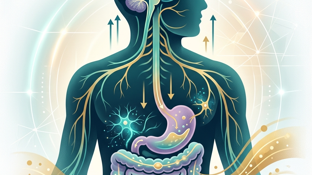
The vagus nerve serves as a major bidirectional highway between the brain and the gastrointestinal tract, carrying signals that influence how food moves through the system, how secretions are released, and how the gut communicates satiety or discomfort back to central regulatory centers. This highway operates through myelinated and unmyelinated fibers that transmit both rapid electrical impulses and slower modulatory chemical signals, allowing the brain to fine-tune digestion in real time while the gut reports back on nutrient density, microbial activity, and mechanical stretch. Understanding this connection matters because digestive comfort, nutrient absorption, and even mood states often trace back to the same cranial nerve that also modulates heart rate and breathing. For example, after eating a fiber-rich meal, vagal afferents may register gradual distension in the ileum and signal the brainstem to adjust subsequent gastric emptying speed, illustrating how one pathway integrates physical sensations with regulatory responses. Readers will encounter the anatomical layout, the physiological mechanisms at work during a meal, the patterns research has identified, and the kinds of daily practices some people explore to support vagal signaling.
Because the vagus nerve is the longest cranial nerve and the primary parasympathetic outflow to the viscera, its activity can shift the digestive tract between states of active processing and relative quiet. When tone is adequate, the nerve tends to promote coordinated peristalsis, appropriate gastric acid and enzyme release, and efficient communication along the gut-brain axis. When signaling is reduced, transit may slow or secretions may become less synchronized, experiences many individuals describe as heaviness, irregular bowel patterns, or reduced appetite awareness. This article examines these relationships through anatomy, physiology, and available evidence without offering individualized recommendations. Everyday scenarios, such as noticing slower digestion during periods of high work stress versus smoother transit on relaxed weekends, highlight how autonomic shifts manifest in tangible ways that people can observe without specialized equipment.
How the Vagus Nerve Works
The vagus nerve originates in the medulla oblongata and descends through the neck, thorax, and abdomen, sending branches to the heart, lungs, and nearly the entire gastrointestinal tract from the esophagus to the proximal colon. Its parasympathetic fibers release acetylcholine at target organs, slowing heart rate, stimulating bronchial constriction, and increasing gut motility and glandular secretion. This “rest-and-digest” profile stands in contrast to sympathetic activation, which generally inhibits digestive processes to redirect resources elsewhere. Because roughly 80 percent of vagal fibers are afferent, the nerve also relays continuous sensory information from the gut wall, liver, and pancreas back to the brainstem, forming a core component of the gut-brain axis. In practice, this means that after consuming a meal containing both protein and fat, stretch receptors in the stomach wall activate vagal afferents that travel upward while simultaneously triggering local acetylcholine release to sustain mixing contractions.
Heart-rate variability, particularly the high-frequency component, serves as one noninvasive window into vagal tone. Greater variability between successive heartbeats often reflects stronger parasympathetic influence via the vagus, while lower variability can accompany reduced vagal outflow. The same nerve that contributes to these cardiac rhythms also modulates gastrointestinal smooth muscle and enteric neurons, illustrating how a single cranial nerve participates in both cardiovascular regulation and digestion. Research on cardiac vagal tone therefore supplies indirect clues about vagal capacity that may extend to visceral organs. Someone tracking daily HRV with a wearable device might notice that an evening walk elevates variability readings the next morning, coinciding with reports of less post-meal bloating, though such correlations remain observational and vary widely between people.
The nerve’s extensive branching also allows it to influence the enteric nervous system, sometimes called the “second brain” of the gut. Vagal afferents detect mechanical stretch, nutrient composition, and inflammatory signals within the intestinal lumen, then transmit this data to the nucleus tractus solitarius. From there, efferent commands can adjust motility patterns or trigger local reflexes such as the gastrocolic response. This closed-loop communication helps explain why changes in breathing rate or posture, both of which engage vagal pathways, sometimes coincide with shifts in digestive comfort. For instance, adopting a seated forward fold after dinner can gently compress abdominal contents, prompting vagal afferents to signal for increased peristalsis that moves contents along the colon without requiring voluntary effort.
The Vagus Nerve’s Role in Digestive Regulation
Within the stomach, vagal efferents stimulate parietal cells to secrete hydrochloric acid and chief cells to release pepsinogen while also relaxing the proximal stomach to accommodate a meal without excessive pressure rise. These coordinated actions rely on acetylcholine acting on muscarinic receptors and on local release of gastrin and histamine that amplify the vagal signal. When vagal drive is lower, acid output may decline and gastric accommodation can become less efficient, patterns some people notice as early fullness or prolonged feelings of heaviness after eating. The same fibers continue into the small intestine, where they promote peristaltic waves that mix chyme with pancreatic enzymes and bile. A concrete example occurs when someone eats a mixed salad with olive oil dressing: initial vagal stimulation encourages receptive relaxation so the stomach can hold the volume, followed by graded acid release calibrated to the fat content detected by duodenal sensors.
Further along the tract, vagal afferents monitor nutrient arrival and mechanical distension, sending graded signals that help terminate meals and initiate the migrating motor complex during fasting periods. This feedback loop contributes to the sense of satisfaction after eating and to the timing of subsequent hunger. Disruption in afferent traffic can therefore alter both appetite regulation and the rhythm of interdigestive motility, experiences individuals sometimes describe as unpredictable hunger cues or irregular bowel movements. The nerve’s influence also extends to enteroendocrine cells that release hormones such as cholecystokinin and glucagon-like peptide-1, linking mechanical and chemical sensing into a unified regulatory network. In daily life this might appear as feeling comfortably full after a balanced lunch yet experiencing renewed hunger signals two hours later if prior vagal feedback was dampened by rushed eating.
Pancreatic exocrine secretion and gallbladder contraction receive vagal input as well, ensuring that digestive enzymes and bile arrive in proportion to the nutrient load entering the duodenum. When these signals are dampened, fat digestion in particular may proceed more slowly, occasionally producing sensations of bloating or loose stools after rich meals. At the same time, vagal anti-inflammatory pathways can modulate intestinal permeability and immune cell activity within the gut wall, illustrating how the nerve participates in both motor-secretory functions and mucosal defense. These overlapping roles mean that changes in vagal tone rarely affect only one aspect of digestion; instead, motility, secretion, and barrier integrity tend to shift together. A person who routinely eats late at night may observe that the combination of reduced vagal tone from circadian misalignment and higher fat intake leads to next-day reports of sluggish transit.
Many people report that periods of lower stress or improved sleep coincide with more predictable digestion, consistent with the nerve’s sensitivity to overall autonomic balance. Conversely, sustained sympathetic dominance can reduce vagal outflow to the gut, slowing transit and altering secretion patterns. Because the same afferent fibers also project to brainstem centers that influence mood and alertness, digestive sensations and emotional tone often travel along shared pathways, though the direction of influence varies between individuals and situations. Nuance arises here because acute stress might suppress appetite via vagal withdrawal, whereas chronic low-level tension can produce the opposite pattern of irregular cravings once the stressor subsides.
What the Research Shows
Studies of the gut-brain axis highlight the vagus nerve as a primary anatomical substrate for bidirectional communication between the gastrointestinal tract and the central nervous system. Vagus nerve as modulator of the brain–gut axis reviews how vagal sensory neurons convey microbial and nutrient signals that can alter brainstem activity and, in turn, descending efferent tone. Complementary work on vagal sensory neurons and gut–brain signaling details the molecular receptors these neurons express, including those sensitive to short-chain fatty acids and mechanical stretch, providing a mechanistic basis for observed links between intestinal content and vagal firing rates. Laboratory models demonstrate that selective activation of these receptors produces measurable changes in brainstem neuronal firing within seconds, offering a window into how dietary fiber fermentation might influence vagal traffic over longer periods.
Human evidence also connects vagal pathways to measurable digestive outcomes. Cleveland Clinic’s overview of vagus nerve function notes that the nerve supplies parasympathetic innervation to the stomach and intestines, directly supporting motility and secretion. Neuroanatomical mapping in NIH StatPearls on cranial nerve 10 confirms the extensive abdominal branching pattern that enables these effects. Meanwhile, research on heart rate variability and cardiac vagal tone demonstrates that higher vagal indices often accompany better-coordinated autonomic regulation, a pattern that may extend to gastrointestinal function even though direct gut-HRV correlations remain an active area of study. Longitudinal observations in healthy volunteers show that individuals with higher baseline variability tend to report more consistent bowel habits, although confounding lifestyle factors prevent causal claims.
Additional investigations into vagal stimulation and visceral function suggest that enhancing afferent or efferent traffic can influence gastric emptying rates and intestinal transit in controlled settings. Work examining vagus nerve stimulation, sleep-disordered breathing, and sleep quality indirectly supports the broader role of vagal tone in autonomic balance, because restorative sleep itself is associated with improved digestive regularity in observational data. Together these sources portray the vagus nerve as an integrative conductor rather than a single on-off switch, with effects that scale according to the nerve’s overall traffic volume and the health of its target organs. Researchers continue to explore dose-response relationships, noting that brief versus prolonged stimulation protocols produce distinct temporal patterns of motility change.
Practical Ways to Support Your Vagus Nerve
- Slow, extended exhales—lengthening the out-breath relative to the in-breath can increase high-frequency heart-rate variability, a marker often linked to greater vagal outflow that reaches the digestive organs.
- Humming or gentle gargling—vibration of the vocal cords and pharynx stimulates vagal branches in the neck, providing a simple mechanical input that some people incorporate before or after meals.
- Gentle cold exposure, such as splashing cool water on the face—activates the diving reflex, which engages vagal pathways and may produce a transient shift toward parasympathetic dominance.
- Paced breathing at approximately six breaths per minute—synchronizes heart-rate oscillations with respiratory rhythm, often elevating vagal tone measures that correlate with broader autonomic regulation.
- Light movement such as walking after eating—promotes mechanical and neural signals that complement vagal control of intestinal motility without requiring intense effort.
- Consistent morning light exposure combined with steady sleep timing—helps entrain circadian rhythms that influence both vagal activity and gastrointestinal hormone release across the day.
When to Talk to a Professional
Sudden, severe abdominal pain, persistent vomiting, unexplained weight loss, or blood in the stool warrant prompt medical evaluation regardless of any interest in vagal pathways. These symptoms can signal conditions that require diagnostic clarification and targeted care. Similarly, difficulty swallowing, chronic reflux that does not respond to basic adjustments, or alternating severe constipation and diarrhea should be assessed by a clinician experienced in gastrointestinal disorders. Early evaluation allows differentiation between functional patterns and structural concerns that may need imaging or laboratory follow-up.
Changes in digestion that coincide with chest pain, dizziness, or shortness of breath also merit immediate attention, because cardiac or pulmonary issues can overlap with vagal symptoms. Individuals who notice progressive difficulty with swallowing or new-onset hoarseness alongside digestive changes may benefit from an ear-nose-throat or neurology consultation to examine vagal motor function directly. Professional guidance ensures that any underlying structural or inflammatory processes receive appropriate investigation. Timing matters here, as intermittent versus progressive symptoms often point toward different investigative priorities.
Common Questions
How quickly can vagal tone change with simple breathing practices?
High-frequency heart-rate variability can shift measurably within minutes of slow breathing, yet sustained alterations in baseline tone generally develop over weeks of consistent practice as autonomic balance recalibrates. Individual responses vary with age, fitness, and concurrent health factors. Some people observe that a single five-minute session of extended exhales produces an immediate sense of abdominal relaxation, while others require repeated daily exposure before noticing changes in post-meal comfort.
Does poor digestion always indicate low vagal tone?
Digestive difficulties can arise from many sources, including diet composition, microbial balance, medications, and structural issues. Reduced vagal signaling represents one possible contributor rather than a universal explanation, which is why professional assessment remains important when symptoms persist. For example, lactose intolerance can produce bloating irrespective of vagal status, underscoring the value of isolating variables through structured observation or testing.
Can vagal pathways be overstimulated?
Excessive parasympathetic activation can produce bradycardia or excessive gastric acid in susceptible individuals, though such responses are uncommon with the gentle daily practices described earlier. Monitoring personal comfort and seeking medical advice for unusual symptoms helps maintain safety. Nuance lies in recognizing that what feels calming to one person, such as prolonged cold-water exposure, may trigger dizziness in another, illustrating the importance of individualized pacing.
Is there a single best practice for everyone?
No universal protocol exists; responses differ according to age, health status, and daily routines. Beginning with the least demanding options, such as extended exhales or consistent sleep timing, allows individuals to observe effects before adding further elements. Tracking subjective digestion alongside simple metrics like morning energy can reveal personal patterns without requiring advanced equipment.
How does the gut microbiome interact with vagal signaling?
Microbial metabolites can activate vagal afferents, which in turn influence brainstem centers that adjust efferent tone back to the gut. This bidirectional loop means that dietary patterns supporting microbial diversity may indirectly affect vagal traffic, though direct causation requires further study. Fermentable fibers that yield short-chain fatty acids provide one everyday illustration, as their breakdown products engage receptors on vagal endings that subsequently modulate motility timing.
The vagus nerve’s extensive reach into digestive physiology underscores why attention to autonomic balance can intersect with everyday gut experiences. By appreciating the nerve’s anatomical layout, its motor and sensory roles, and the evidence that continues to accumulate, readers gain a clearer framework for noticing patterns without expecting any single practice to resolve complex symptoms. Continued professional partnership remains the appropriate route whenever digestive concerns intensify or persist.
Have a question?
Have a question about something specific? Send us a message.
Visit VagusSkool.com/contact — we'll try to get back to you within 24 hours.