The Vagus Nerve: How It Shapes Sleep Quality, Stress Recovery, and Social Connection
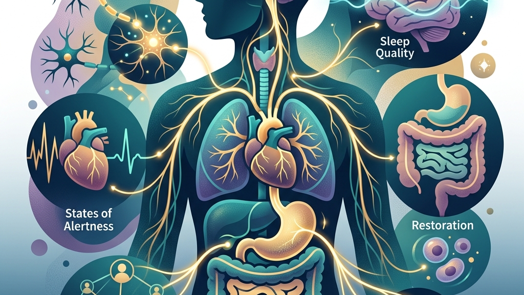
How the vagus nerve works
The vagus nerve, designated cranial nerve X, emerges from the medulla oblongata and extends through the neck, chest, and abdomen, innervating the heart, lungs, esophagus, stomach, intestines, and other structures. Its two main trunks carry both motor and sensory fibers, allowing bidirectional communication that supports parasympathetic control—the branch of the autonomic nervous system associated with conservation of energy and restoration. Unlike the sympathetic system’s rapid mobilization for action, vagal activation tends to slow heart rate, promote gastrointestinal motility, and dampen inflammatory signaling through the cholinergic anti-inflammatory pathway. A key feature of vagal function appears in its modulation of heart-rate variability, the moment-to-moment fluctuation in intervals between heartbeats. Higher variability often reflects stronger vagal influence, indicating an intact “vagal brake” that can be released during demand and reapplied during safety. This brake operates through the nucleus ambiguus and dorsal motor nucleus, adjusting cardiac output without requiring conscious effort. When vagal tone is robust, transitions between activity and rest occur more smoothly; when it is reduced, the system may linger in higher sympathetic tone even during periods that would otherwise favor recovery. The gut-brain axis further illustrates the nerve’s integrative role. Roughly 80 percent of vagal fibers transmit information upward from visceral organs to the brainstem, conveying mechanical stretch, nutrient presence, and microbial metabolites. These ascending signals reach areas such as the nucleus tractus solitarius, which then projects to higher centers involved in mood and arousal. Research on vagal sensory neurons shows they detect specific gut-derived molecules and relay them rapidly, influencing both local reflexes and broader brain states that affect sleep onset and emotional regulation. Because the nerve also supplies laryngeal and pharyngeal muscles, its activity participates in voice production and swallowing. Subtle changes in vagal outflow can therefore alter vocal tone and resonance, features that listeners unconsciously register during conversation. This anatomical arrangement links internal regulatory processes with external signaling, providing a physiological substrate for the interplay between bodily state and social behavior.Vagal Tone and Sleep Architecture
During the transition into sleep, vagal activity rises to support the parasympathetic dominance required for sustained non-REM stages. Slowed heart rate, reduced respiratory rate, and increased digestive activity all depend on adequate vagal outflow; when this outflow is insufficient, cortical arousal may persist and fragment sleep continuity. The nerve’s influence extends into REM periods as well, where it helps stabilize autonomic fluctuations that otherwise could trigger micro-arousals or shifts in muscle tone. Heart-rate variability measured during nighttime rest often serves as a proxy for vagal contribution to sleep maintenance. Higher nocturnal HRV correlates with longer duration of slow-wave sleep and fewer awakenings, consistent with the nerve’s role in lowering sympathetic drive. Conversely, conditions that impair vagal signaling—such as chronic inflammation or mechanical compression along its course—can coincide with lighter sleep and more frequent stage shifts, even when total sleep time appears adequate on paper. Many individuals notice that evenings marked by easy breathing and a sense of abdominal relaxation precede more consolidated sleep, while evenings of shallow breathing or digestive discomfort precede restless nights. These subjective reports align with the nerve’s dual sensory and motor functions: ascending signals from a calm gut reinforce brainstem sleep-promoting circuits, while descending motor signals relax laryngeal and pharyngeal tone without collapsing the airway. Over successive nights, small improvements in vagal regulation may accumulate into measurable differences in morning refreshment and daytime alertness. The relationship is bidirectional. Poor sleep itself can blunt vagal responsiveness the following day, creating a cycle in which reduced nighttime vagal tone predicts lower daytime HRV and vice versa. This loop underscores why interventions targeting vagal pathways often address both sleep timing and daytime regulatory capacity rather than sleep duration alone.Stress Recovery and the Vagal Brake
After an acute stressor, the vagus nerve participates in returning cardiovascular, respiratory, and immune parameters toward baseline. The “vagal brake” refers to the rapid re-engagement of parasympathetic outflow that lowers heart rate and restores heart-rate variability once the threat has passed. Efficient braking depends on intact afferent feedback from baroreceptors and visceral organs; without it, sympathetic activation may decay slowly, leaving residual muscle tension, elevated breathing rate, and narrowed attentional focus. Mechanistically, vagal stimulation inhibits sympathetic preganglionic neurons in the spinal cord and dampens hypothalamic-pituitary-adrenal axis activity through projections to the paraventricular nucleus. This inhibition reduces circulating catecholamines and supports resolution of inflammatory cascades initiated during the stress response. When vagal tone is low, the resolution phase lengthens, increasing the cumulative physiological cost of repeated stressors over hours or days. People commonly describe a lingering sense of vigilance or digestive unease after demanding events, even when the original trigger has ended. These sensations correspond to delayed vagal reactivation: heart rate remains elevated, breathing stays thoracic rather than diaphragmatic, and gastrointestinal motility remains suppressed. Over time, repeated incomplete recovery can lower the threshold for future stress responses, making the brake harder to reapply. Because vagal afferents also convey information about bodily state back to the brain, successful stress recovery often involves noticing subtle shifts—such as warmth returning to the hands or a deeper sigh occurring spontaneously. These interoceptive cues reflect restored vagal traffic and can themselves reinforce further parasympathetic engagement through ascending pathways to regulatory centers in the brainstem and forebrain.Social Connection and Vagal Pathways
Social engagement recruits vagal circuits through both motor and sensory routes. The ventral vagal complex, centered around the nucleus ambiguus, supplies the muscles of the face, middle ear, larynx, and pharynx that produce expressive vocal prosody and detect human voices against background noise. When this system is active, individuals tend to exhibit warmer vocal timbre, more varied intonation, and spontaneous facial expressions that signal safety to others. Physiological safety cues travel in both directions. A calm physiological state increases the likelihood of emitting such cues, while receiving them from others can further increase vagal tone via afferent feedback. This feedback loop helps explain why conversations in a low-threat environment often deepen into slower breathing and greater abdominal relaxation, whereas tense exchanges maintain higher muscle tone in the throat and chest. Research on vagal sensory neurons indicates they also participate in sensing affiliative touch and proximity, relaying these signals to brainstem areas that modulate arousal. Consequently, moments of genuine connection may coincide with transient increases in heart-rate variability and reductions in inflammatory markers, effects that accumulate when positive social contact occurs regularly. Reduced vagal tone, by contrast, can narrow the range of perceived safety, making neutral social situations feel more effortful or less rewarding. Voice and throat sensations frequently serve as early indicators of shifting vagal state during social interaction. A sudden tightening or flattening of vocal resonance may reflect momentary withdrawal of ventral vagal influence, while a return of natural resonance often accompanies renewed ease. These micro-shifts operate below conscious awareness for most people yet contribute to the felt difference between interactions that leave one energized and those that leave one drained.What the research shows
Multiple lines of evidence link vagal function to the domains discussed. Cleveland Clinic summaries of vagus-nerve anatomy emphasize its extensive parasympathetic distribution and role in heart-rate regulation, providing a foundation for understanding HRV changes during sleep and recovery. NIH StatPearls on cranial nerve 10 detail the nerve’s brainstem nuclei and laryngeal branches, clarifying how motor output supports both airway patency during sleep and vocal prosody during social exchange. Studies examining vagus-nerve stimulation in relation to sleep-disordered breathing report associations between enhanced vagal signaling and improved sleep continuity, particularly through effects on respiratory control and arousal thresholds. Parallel work on heart-rate variability and cardiac vagal tone demonstrates that higher resting HRV predicts faster cardiovascular recovery after laboratory stressors and correlates with self-reported calmness in daily life. Investigations into the vagus nerve as modulator of the brain–gut axis highlight bidirectional signaling that influences both gastrointestinal comfort and central arousal states relevant to sleep initiation. Additional findings from research on vagal sensory neurons and gut–brain signaling show that specific populations of vagal afferents respond to mechanical and chemical stimuli in the gut, relaying information that can either promote or disrupt sleep depending on the state of the viscera. Across these sources, the pattern is consistent: vagal integrity supports smoother transitions among sleep, recovery, and social engagement, while disruptions in vagal traffic correspond to greater fragmentation in each domain. Effect sizes vary by population and measurement method, and individual responses remain heterogeneous.Practical ways to support your vagus nerve
- Slow, extended exhales—lengthening the out-breath to roughly twice the duration of the in-breath—can increase vagal cardioinhibitory outflow within a single session by enhancing baroreceptor stimulation.
- Humming or gentle gargling engages laryngeal branches of the vagus, providing motor activation that may reinforce ventral vagal tone and alter middle-ear muscle tension linked to social sound processing.
- Brief, tolerable cold exposure such as cool water on the face or neck stimulates vagal afferents via the diving reflex, often producing an immediate shift toward lower heart rate and greater heart-rate variability.
- Paced breathing at approximately six breaths per minute aligns with resonance frequency for many adults, amplifying vagal oscillations measurable in HRV recordings.
- Light movement that incorporates rhythmic locomotion, such as walking at a comfortable pace, couples respiratory and cardiac rhythms in ways that can support vagal reactivation after sedentary periods.
- Consistent morning light exposure combined with stable sleep timing helps entrain brainstem nuclei that regulate both circadian arousal and nocturnal vagal dominance.
When to talk to a professional
Persistent difficulty falling or staying asleep, sudden changes in heart rhythm, unexplained digestive distress, or marked social withdrawal that interferes with daily functioning warrant evaluation by a qualified clinician. These experiences can stem from multiple physiological or neurological factors, and self-directed practices are not substitutes for assessment when symptoms are severe, progressive, or accompanied by chest pain, shortness of breath, or mood changes that feel overwhelming. A medical professional can determine whether further testing of autonomic function or other systems is appropriate.Common questions
Does vagal tone change quickly or does it require months of practice?
Acute shifts in vagal activity can occur within minutes through breathing or posture adjustments, yet sustained improvements in baseline tone typically reflect repeated, consistent engagement over weeks rather than isolated efforts.
Can HRV measurements at home reliably indicate vagal function?
Consumer devices provide directional information about heart-rate variability, but they do not isolate vagal contributions from other autonomic influences and should not replace clinical interpretation when symptoms are present.
Is there a single best practice for supporting the vagus nerve?
No universal hierarchy exists; different individuals respond to different entry points such as breath, voice, or movement, and the most useful practice is often the one that can be performed regularly without strain.
Does age affect how readily vagal tone responds to these approaches?
Age-related changes in autonomic flexibility occur, yet research indicates that older adults can still demonstrate measurable HRV increases with targeted breathing or light activity, though the magnitude and speed of change may differ from younger cohorts.
The vagus nerve offers a tangible physiological thread connecting nightly rest, the capacity to settle after pressure, and the felt quality of relating to others. Attending to its signals through ordinary daily rhythms does not eliminate life’s demands, yet it can enlarge the margin within which recovery and connection become more accessible.Have a question?
Have a question about something specific? Send us a message.
Visit VagusSkool.com/contact — we'll try to get back to you within 24 hours.