The Vagus Nerve, Restorative Sleep, and the Physiology of Breath and Stress Recovery
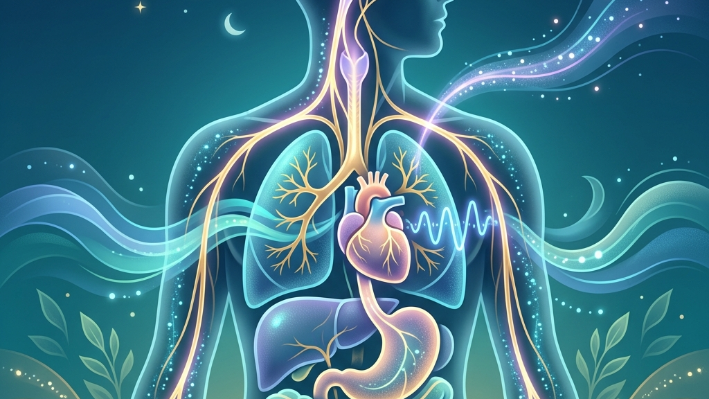
How the vagus nerve works
The vagus nerve, designated as cranial nerve X, originates in the medulla oblongata and extends bilaterally through the neck, thorax, and abdomen. Its sensory and motor fibers form the longest cranial nerve, carrying afferent signals from organs back to the brainstem and efferent signals outward to regulate organ function. In plain terms, this nerve constitutes the main parasympathetic highway, slowing heart rate, promoting digestive motility, and dampening inflammatory responses when active. Afferent fibers vastly outnumber efferent ones, meaning the nerve primarily reports visceral states upward rather than issuing commands downward. This arrangement supports the gut-brain axis, where microbial metabolites, mechanical stretch in the intestines, and local immune activity generate signals that travel via vagal afferents to influence brainstem nuclei and higher cortical areas involved in mood and arousal. Research on this axis highlights how vagal transmission integrates metabolic and immune information with central autonomic control. For instance, after a meal containing fermentable fibers, short-chain fatty acids produced by gut bacteria bind to receptors on enteroendocrine cells; these cells then release signaling molecules that activate nearby vagal endings, which relay the information through the nucleus tractus solitarius to adjust both local gut motility and distant cortical areas that influence perceived energy levels later in the day. Heart-rate variability arises largely from vagal modulation of the sinoatrial node. Respiratory sinus arrhythmia, the natural fluctuation in beat-to-beat intervals synchronized with breathing, reflects vagal tone in real time. Higher baseline variability generally corresponds to greater parasympathetic dominance, allowing the system to shift flexibly between arousal and rest. The vagus nerve thus functions as a dynamic brake on sympathetic acceleration, releasing during exhalation and engaging more strongly during states of safety or recovery. In everyday terms, this brake can be observed when someone sits quietly after climbing stairs: the interval between heartbeats lengthens noticeably on each out-breath, an effect mediated by acetylcholine release at the sinoatrial node that temporarily hyperpolarizes pacemaker cells and slows the next depolarization.Sleep and Vagal Tone
During non-REM sleep stages, parasympathetic dominance increases and vagal outflow to the heart and viscera rises, supporting slower heart rates and reduced metabolic demand. This shift facilitates deeper slow-wave activity, which in turn correlates with enhanced vagal afferent feedback from the gut and cardiovascular system. The resulting loop helps consolidate memory and clear metabolic byproducts, processes that rely on stable autonomic balance rather than persistent sympathetic activation. Slow-wave sleep, in particular, features large-amplitude delta oscillations generated by synchronized cortical firing; these oscillations coincide with heightened vagal traffic from baroreceptors that detect the modest nocturnal drop in blood pressure, creating a feedback loop in which lower cardiac output further reinforces parasympathetic dominance. Vagal sensory neurons detect changes in blood pressure, lung inflation, and gastrointestinal tone that occur across sleep cycles. When these signals remain within a narrow, regulated range, transitions between sleep stages occur more smoothly and awakenings decrease. Disruption of vagal signaling, whether from chronic inflammation or mechanical factors, can fragment sleep continuity by allowing sympathetic surges to intrude during periods when parasympathetic tone should predominate. Consider an individual who consumes a large, late meal: gastric distension activates mechanoreceptors whose signals ascend via the vagus; if the volume or composition delays gastric emptying, the sustained afferent barrage can raise the threshold for entering deeper sleep stages, resulting in more frequent micro-arousals visible on a home sleep tracker as brief spikes in heart rate. Many people notice that nights following days of lower stress or consistent meal timing coincide with fewer nighttime arousals and a subjective sense of deeper rest. Conversely, evenings marked by digestive discomfort or elevated heart rate often precede lighter, more interrupted sleep. These patterns align with the nerve’s role in relaying visceral calm or unrest to brainstem centers that gate sleep depth and duration. The interplay extends to morning wakefulness. A robust nocturnal rise in vagal activity tends to leave residual parasympathetic tone that supports a gradual rather than abrupt cortisol increase upon waking. Individuals who track subjective energy often describe steadier morning alertness when preceding sleep periods included minimal sympathetic intrusions, consistent with preserved vagal modulation throughout the night. Over successive nights, this pattern can be influenced by the timing of last food intake relative to bedtime, because vagal afferents from the duodenum continue to transmit nutrient-sensing signals for several hours after the final meal.Resting Heart Rate and the Vagal Brake
The vagal brake refers to tonic parasympathetic inhibition of the heart that keeps resting rate below the intrinsic pacemaker rhythm. During stress recovery, this brake re-engages after sympathetic activation subsides, producing a measurable deceleration in heart rate and a rebound in heart-rate variability. The speed and completeness of this re-engagement depend on intact vagal efferent pathways and adequate afferent feedback indicating that the threat has passed. In laboratory settings, researchers measure this re-engagement by tracking the time required for heart-rate variability to return to 90 percent of pre-stressor baseline after a standardized mental arithmetic task; shorter recovery times correlate with stronger baseline vagal outflow from the nucleus ambiguus. Stress-recovery physiology therefore hinges on the vagus nerve’s capacity to override residual sympathetic tone. When afferent signals from baroreceptors and visceral organs confirm safety, medullary cardioinhibitory neurons increase firing, releasing acetylcholine onto the sinoatrial node. This mechanism lowers oxygen demand and supports anabolic processes such as tissue repair and digestion that were suppressed during the stress response. A concrete illustration occurs after an unexpected work deadline: once the immediate pressure lifts, an individual may notice the pulse dropping from 95 to 72 beats per minute over roughly eight minutes while seated, accompanied by a subtle warming sensation across the upper chest as cutaneous blood flow increases under renewed parasympathetic control. Observations commonly reported include a gradual calming of pulse and breathing after a demanding event, sometimes accompanied by a sensation of warmth in the chest or abdomen. These subjective shifts coincide with the physiological window in which vagal tone reasserts dominance, typically within minutes to hours depending on the intensity and duration of the preceding stressor. Prolonged delays in this return often accompany sustained muscle tension or gastrointestinal unease. Over repeated cycles, the efficiency of vagal re-engagement shapes how resilient the system appears to recurring demands. Efficient braking reduces the cumulative load on cardiovascular and immune tissues, whereas sluggish return leaves residual sympathetic bias that can blunt subsequent recovery periods. The vagus nerve thus acts as both sensor and effector in the daily oscillation between mobilization and restoration.Voice, Throat, and the Vagus Nerve
Motor fibers of the vagus nerve innervate the muscles of the larynx and pharynx, directly controlling vocal-fold tension, swallowing, and airway protection. These same fibers participate in respiratory rhythm generation through connections with the nucleus ambiguus, allowing breath patterns to influence and be influenced by vagal outflow. Slow, rhythmic breathing therefore engages laryngeal and pharyngeal structures that send afferent traffic back along the nerve, reinforcing parasympathetic tone. The recurrent laryngeal branch, for example, supplies motor innervation to all intrinsic laryngeal muscles except the cricothyroid; its activity modulates vocal-fold length and tension on a millisecond timescale, which in turn alters subglottal pressure and feeds back to medullary respiratory centers. During exhalation, especially when prolonged, vagal cardiac fibers increase their inhibitory effect on heart rate, producing the expiratory component of respiratory sinus arrhythmia. This mechanical linkage means that breath practices altering airway resistance or duration can measurably shift heart-rate variability within a single cycle. The throat thus serves as both an output pathway for vagal motor signals and an input site for sensory feedback that calibrates autonomic state. People frequently observe that deliberate slowing of the out-breath or the production of low-frequency vocal sounds coincides with a perceptible drop in heart rate and a sense of throat or chest relaxation. These sensations align with increased vagal afferent discharge from stretch receptors in the lungs and larynx. Conversely, rapid or shallow breathing tends to reduce this feedback, leaving heart-rate variability lower and sympathetic tone relatively higher. The bidirectional relationship extends beyond deliberate practice. Spontaneous sighing, yawning, and even quiet humming recruit the same laryngeal vagal pathways, providing brief windows of heightened parasympathetic activity. Over time, consistent patterns of breathing that emphasize extended exhalation appear to support more stable baseline vagal tone, though individual responses vary with overall health status and concurrent autonomic load.What the research shows
Studies of heart-rate variability consistently link higher vagal tone, indexed by respiratory sinus arrhythmia and other time-domain measures, to improved cardiac autonomic flexibility. Heart Rate Variability and Cardiac Vagal Tone reviews evidence that vagal modulation buffers against excessive sympathetic drive and supports recovery after physical or psychological challenge. Sleep research has examined vagus nerve stimulation parameters in relation to sleep-disordered breathing and overall sleep quality metrics. Vagus Nerve Stimulation, Sleep-Disordered Breathing & Sleep Quality summarizes findings indicating that targeted stimulation can influence upper-airway muscle tone and arousal thresholds, though results differ across stimulation protocols and patient populations. Anatomical tracing and functional imaging studies map the vagus nerve’s role in gut–brain communication. Vagus Nerve as Modulator of the Brain–Gut Axis and Vagal Sensory Neurons and Gut–Brain Signaling detail how vagal afferents convey nutrient, microbial, and inflammatory signals that reach brainstem and limbic structures, thereby influencing both visceral regulation and central arousal systems relevant to sleep and stress. Basic anatomical references confirm the nerve’s extensive distribution and mixed sensory-motor composition. Vagus Nerve: Function, Location & Conditions and Neuroanatomy, Cranial Nerve 10 (Vagus Nerve) provide overviews of its course and the parasympathetic effects that underpin the mechanisms discussed above.Practical ways to support your vagus nerve
- Slow extended exhales performed for several minutes can increase the duration of vagal cardiac inhibition within each breath cycle by lengthening the expiratory phase that naturally augments parasympathetic outflow.
- Humming or gentle gargling activates laryngeal motor fibers of the vagus and generates low-frequency vibrations that travel along afferent pathways, often producing a rapid subjective calming effect.
- Gentle cold exposure, such as cool water on the face, stimulates trigeminal and vagal afferents that can trigger a brief parasympathetic rebound observable as reduced heart rate.
- Paced breathing at approximately six breaths per minute aligns respiratory frequency with the resonance frequency of the baroreflex, amplifying heart-rate variability mediated by the vagus nerve.
- Light movement such as walking after meals promotes mechanical stimulation of gastrointestinal vagal afferents through peristalsis and posture changes, supporting postprandial parasympathetic dominance.
- Morning light exposure combined with consistent sleep timing helps entrain circadian rhythms that in turn influence nocturnal vagal tone and daytime autonomic recovery capacity.
When to talk to a professional
Sudden or severe changes in heart rhythm, breathing difficulty, persistent insomnia, or unexplained gastrointestinal symptoms warrant prompt medical evaluation to rule out underlying conditions. These signs may reflect issues beyond normal autonomic variation and require assessment by a qualified clinician. Individuals experiencing chest pain, dizziness on standing, or rapid unexplained weight changes should seek professional guidance rather than attempting to interpret these through an autonomic lens alone. Early consultation helps distinguish benign fluctuations from those needing targeted investigation.Common questions
How quickly can vagal tone change?
Beat-to-beat adjustments occur within a single respiratory cycle, while longer-term shifts in baseline tone develop over days to weeks with consistent patterns of breathing, sleep, and movement.
Does age affect vagal function?
Resting vagal tone tends to decline gradually with advancing age, yet the nerve’s responsiveness to respiratory and postural inputs remains modifiable across the lifespan in most people.
Can digestive issues influence sleep through the vagus nerve?
Vagal afferents carry signals from the gut that reach brainstem sleep-regulatory centers, so gastrointestinal discomfort can increase arousal probability during vulnerable sleep stages.
Is heart-rate variability the only marker of vagal tone?
While widely used, heart-rate variability captures only cardiac vagal modulation; laryngeal, gastrointestinal, and inflammatory pathways also reflect vagal activity and may not move in perfect synchrony.
The vagus nerve integrates information from the heart, lungs, and digestive tract into a continuous stream that shapes how sleep unfolds, how quickly stress physiology resolves, and how breath patterns either support or hinder return to baseline. Attention to these mechanisms offers a coherent perspective on autonomic flexibility without promising uniform results. Consistent observation of personal patterns, paired with professional care when symptoms intensify, remains the most reliable approach to understanding one’s own physiological responses.Have a question?
Have a question about something specific? Send us a message.
Visit VagusSkool.com/contact — we'll try to get back to you within 24 hours.