The Vagus Nerve’s Influence on Sleep Architecture and Stress Recovery Pathways
How the Vagus Nerve Works
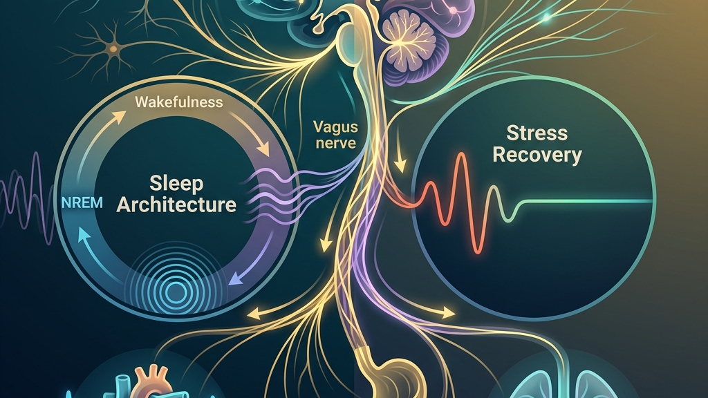
The vagus nerve, designated as cranial nerve X, emerges from the medulla oblongata and extends through the neck, thorax, and abdomen, forming the longest cranial nerve pathway. It carries both efferent fibers that transmit signals from the brain to peripheral organs and afferent fibers that relay sensory information back to the brainstem. This bidirectional traffic allows the nerve to modulate heart rate, respiratory rhythm, and gastrointestinal motility while simultaneously informing central regulatory centers about the state of the viscera. Efferent fibers originate in the dorsal motor nucleus and nucleus ambiguus, traveling outward to release acetylcholine at target organs and thereby slowing sinoatrial node firing in the heart or increasing peristaltic waves in the stomach and intestines. Afferent fibers, by contrast, gather data from baroreceptors in the aortic arch, chemoreceptors in the lungs, and mechanoreceptors along the gut wall, returning that information through the nodose and jugular ganglia to the nucleus of the solitary tract. The result is a closed-loop system in which, for instance, a sudden rise in blood pressure detected by aortic sensors can trigger an immediate vagal command to reduce heart rate before conscious awareness occurs. In its parasympathetic capacity the vagus nerve promotes reduced heart rate and increased digestive activity, counterbalancing sympathetic activation. Heart-rate variability, particularly the high-frequency component linked to respiratory sinus arrhythmia, serves as a common noninvasive index of vagal cardiac outflow. Greater variability generally reflects stronger parasympathetic influence, although the measure integrates multiple inputs and requires careful interpretation across contexts. During everyday activities such as reading while seated or conversing calmly, the high-frequency oscillations become visible on a simple pulse-oximeter display as rhythmic accelerations during inhalation and decelerations during exhalation. When someone stands up quickly after prolonged sitting, the same system must rapidly adjust to maintain cerebral perfusion; a robust vagal response helps prevent lightheadedness by fine-tuning vascular tone within seconds. Interpretation of any single reading, however, must account for posture, recent caffeine intake, and ambient temperature, all of which alter the same variability metric. The gut-brain axis further illustrates vagal reach. Roughly 80 percent of vagal fibers are afferent, conveying mechanical, chemical, and microbial signals from the intestines to the brainstem. These signals influence brainstem nuclei that in turn adjust autonomic tone, mood-related circuits, and arousal thresholds. Research on vagal sensory neurons underscores their role in transmitting satiety, inflammation, and microbial metabolite information, thereby shaping whole-body regulatory loops. After a large, fiber-rich meal, stretch receptors along the stomach wall increase firing rates that travel via vagal afferents, prompting the brainstem to reduce overall sympathetic drive and facilitate nutrient absorption. Conversely, when intestinal inflammation elevates circulating cytokines, the same afferent pathway can sustain a low-grade increase in heart rate and vigilance, illustrating how a peripheral digestive event can shift central arousal thresholds without any change in external circumstances.Vagal Tone and the Architecture of Sleep
During the descent into non-rapid-eye-movement sleep, parasympathetic dominance mediated partly by the vagus nerve supports the slowing of heart rate and the consolidation of slow-wave activity. Respiratory patterns become more regular, and the baroreflex operates with heightened sensitivity, allowing blood-pressure fluctuations to be buffered efficiently. Individuals often notice that nights characterized by steadier breathing and lower resting heart rate coincide with feeling more refreshed upon waking, although many additional factors contribute to subjective sleep quality. In practical terms, this means that lying quietly in a darkened room after a consistent wind-down routine allows vagal efferents to predominate, lengthening the interval between successive heartbeats and permitting growth-hormone release during the deepest slow-wave stages. The baroreflex enhancement can be observed indirectly when someone measures overnight pulse and finds that small positional changes produce only brief, quickly corrected pressure shifts rather than sustained elevations. The transition into rapid-eye-movement sleep involves a temporary withdrawal of vagal cardiac control accompanied by sympathetic surges that produce the characteristic variability in heart rhythm. When baseline vagal capacity is robust, these phasic activations remain within narrower bounds and do not provoke prolonged arousals. Studies examining sleep-disordered breathing have observed associations between vagal afferent integrity and the stability of upper-airway muscle tone, suggesting one route by which vagal signaling may influence sleep continuity. For example, a person who maintains higher daytime heart-rate variability through regular low-intensity movement may experience fewer micro-arousals during REM because the airway dilator muscles retain sufficient tone even when brainstem respiratory drive fluctuates. This does not imply that any single habit guarantees uninterrupted sleep, only that the underlying neural substrate participates in the observed stability. Circadian alignment also intersects with vagal function. Morning light exposure and consistent sleep timing help synchronize brainstem clocks that regulate vagal outflow across the 24-hour cycle. People frequently report that maintaining a regular sleep window reduces the latency to sleep onset, an observation consistent with improved parasympathetic recruitment at habitual bedtime. Conversely, irregular schedules can blunt the amplitude of nocturnal vagal modulation, extending the time required for physiological downregulation. Someone who travels across time zones or works rotating shifts often notices that the first night back on a stable schedule brings faster settling, reflecting gradual re-entrainment of medullary oscillators that gate vagal tone. Finally, the gut-brain axis participates in sleep regulation through vagal afferents that carry signals related to microbial metabolites and inflammatory mediators. When intestinal comfort is stable, fewer disruptive afferent volleys reach the brainstem, supporting longer bouts of consolidated sleep. This pathway illustrates how peripheral conditions can influence central sleep processes via the vagus without implying direct causation in every case. A concrete illustration occurs when an individual consumes a late, heavy meal; the resulting mechanical and chemical signals can generate afferent traffic that competes with the normal nocturnal increase in vagal tone, leading to more fragmented early-night sleep even though the person feels tired.Vagal Pathways Supporting Recovery from Stress
After sympathetic activation, the vagus nerve contributes to the re-engagement of parasympathetic control through what is sometimes termed the vagal brake on heart rate. Rapid withdrawal and subsequent reapplication of this brake allow heart rate to return toward resting levels once the stressor subsides. Greater baseline vagal tone, reflected in higher heart-rate variability, is associated in research with more efficient post-stress deceleration, although individual differences in fitness, age, and health status modulate the response. Consider an ordinary scenario in which a driver encounters an unexpected traffic jam and experiences an abrupt sympathetic surge; once the congestion clears, a well-functioning vagal brake produces a measurable deceleration within one or two minutes rather than leaving residual elevation for the remainder of the drive. Respiratory control provides another entry point. Slow, extended exhalations increase vagal efferent traffic to the heart via pulmonary stretch receptors and brainstem integration. This mechanism can be engaged deliberately after demanding events, often producing a perceptible calming of cardiovascular arousal within minutes. Many people describe a shift from a “racing” sensation to a broader sense of settling when such breathing patterns are practiced consistently. The stretch-receptor activation occurs because prolonged exhalation slightly increases intrathoracic pressure, which in turn augments baroreceptor firing that reinforces vagal outflow through the nucleus of the solitary tract. The same afferent pathways that inform the brain about gut state also participate in stress recovery. Inflammatory signals or mechanical tension in the viscera can maintain low-grade sympathetic bias; conversely, conditions that reduce such afferent drive allow vagal efferents to predominate more readily. This bidirectional traffic helps explain why gastrointestinal comfort and cardiovascular settling frequently co-vary during recovery periods. After an acutely stressful meeting, for instance, someone who then sits quietly with an empty stomach may notice both a gradual slowing of heart rate and a reduction in abdominal tension, reflecting simultaneous changes along the shared vagal routes. Over repeated cycles, the cumulative effect of reliable vagal re-engagement after stress appears to support steadier daytime energy and lower nocturnal arousal. Longitudinal observations link higher resting heart-rate variability with reports of feeling less physiologically “on guard” between challenges, yet these associations remain correlational and are shaped by numerous lifestyle and constitutional variables. The pattern suggests that each successful return to baseline may incrementally strengthen the efficiency of the same neural circuits, although the magnitude of change differs across individuals and cannot be isolated from concurrent factors such as physical fitness or social support.What the Research Shows
Neuroanatomical descriptions confirm the vagus nerve’s extensive distribution and mixed afferent-efferent composition, providing the structural basis for its regulatory roles. NIH StatPearls on cranial nerve 10 detail the medullary origins and peripheral branches that enable cardiac, pulmonary, and gastrointestinal modulation. Complementary overviews from Cleveland Clinic on vagus nerve function emphasize its contribution to parasympathetic tone and the clinical relevance of heart-rate variability measures. These foundational accounts map the precise brainstem nuclei and fiber trajectories that allow a single nerve to influence organs distributed across multiple body cavities. Investigations into cardiac vagal tone highlight how respiratory sinus arrhythmia and baroreflex sensitivity index parasympathetic outflow, with implications for both sleep stability and post-stress recovery. PMC article on heart rate variability and cardiac vagal tone reviews the physiological underpinnings and measurement considerations that inform these interpretations. Parallel work on the gut-brain axis demonstrates that vagal afferents convey microbial and inflammatory information capable of influencing brainstem autonomic centers, as summarized in PMC review of the vagus nerve as modulator of the brain–gut axis and PMC article on vagal sensory neurons and gut–brain signaling. The latter studies clarify that metabolites produced by intestinal bacteria can bind to receptors on vagal endings, altering the frequency of action potentials that reach the nucleus of the solitary tract and thereby modulating both autonomic output and arousal-related networks. Specific to sleep, PMC examination of vagus nerve stimulation, sleep-disordered breathing, and sleep quality synthesizes evidence linking vagal pathways to respiratory control during sleep and to overall sleep architecture. These studies collectively illustrate associations rather than isolated causal claims, underscoring the multifactorial nature of both sleep and stress recovery. Across the cited literature, the consistent theme is that vagal signaling participates in multiple regulatory loops whose combined output determines observable physiological states, while the relative contribution of any single loop varies with context and individual physiology.Practical Ways to Support Your Vagus Nerve
- Slow, extended exhales performed for several minutes can increase vagal cardiac outflow by enhancing respiratory sinus arrhythmia, offering a readily accessible entry point that requires no equipment.
- Gentle humming or soft gargling stimulates vagal afferents in the throat and larynx, providing a low-effort sensory input that many find compatible with evening routines.
- Brief, tolerable cold exposure such as cool water on the face or hands engages vagal reflexes via trigeminal and glossopharyngeal pathways, typically producing a transient shift toward parasympathetic dominance.
- Paced breathing at approximately six breaths per minute aligns with the frequency that maximizes heart-rate variability for many adults, allowing practice sessions to be integrated into transitions between activities.
- Light movement such as walking after meals supports vagal gastrointestinal signaling through mechanical and metabolic cues without demanding high exertion.
- Consistent morning light exposure combined with a stable sleep window helps entrain brainstem oscillators that regulate the amplitude of nocturnal vagal tone across successive days.
When to Talk to a Professional
Persistent difficulty falling or staying asleep, sudden changes in resting heart rate, or prolonged physical tension following routine stressors may indicate underlying conditions that extend beyond vagal function alone. Individuals experiencing chest discomfort, severe gastrointestinal symptoms, or marked alterations in consciousness should seek medical assessment promptly. A qualified clinician can evaluate whether autonomic patterns form part of a broader clinical picture and recommend appropriate investigations or interventions. Such evaluation typically includes a detailed history, orthostatic vital-sign measurements, and, when indicated, ambulatory monitoring that captures both daytime activity and nocturnal physiology, allowing any observed vagal metrics to be interpreted within the full clinical context rather than in isolation.Common Questions
Does everyone have the same vagal capacity?
Vagal tone varies with age, fitness level, health status, and prior stress exposure; what registers as typical for one person may differ for another, so comparisons are best avoided. For instance, an endurance-trained adult may display higher average high-frequency heart-rate variability than a sedentary peer of the same age, yet both values can fall within ranges compatible with everyday function when other health parameters remain stable.
Can breathing exercises replace other forms of recovery support?
Breathing practices engage vagal pathways but operate within a larger set of influences including sleep timing, nutrition, and social context; they are best viewed as one accessible tool among many. Their utility appears greatest when integrated with stable schedules and adequate recovery periods rather than used as a standalone intervention.
How long does it take to notice changes in heart-rate variability?
Acute shifts can appear within a single session of slow breathing, while longer-term patterns depend on multiple lifestyle factors and generally require consistent observation over weeks rather than days. Wearable devices that track nightly averages often reveal gradual upward trends only after several weeks of stable routines, underscoring the value of repeated measurement under comparable conditions.
Is there a single best time of day to practice vagal-supportive activities?
Many people find evening practices helpful for sleep onset and morning practices supportive of daytime regulation, yet individual schedules and responses differ and experimentation within safe bounds is common. The key variable appears to be consistency relative to one’s own circadian phase rather than adherence to a universal clock time.
The vagus nerve offers a tangible physiological thread connecting nightly restoration with daytime recovery, inviting continued curiosity about how autonomic balance is cultivated across ordinary days.Have a question?
Have a question about something specific? Send us a message.
Visit VagusSkool.com/contact — we'll try to get back to you within 24 hours.